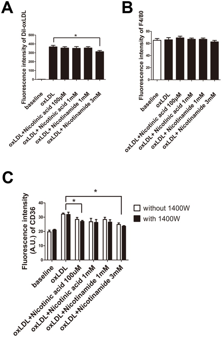
Figure 2. Flow cytometry of Dil-oxLDL, and F4/80.
A. Cells were preincubated with Dil-oxLDL, then Nican in a concentration of 100 µM or 1 mM, or Nicotinamide in a concentration of 1 mM or 3 mM were added. The fluorescence intensity of Dil-oxLDL treated cells was significantly reduced when cell were treated with Nicotinamide at 3 mM. (*, P<0.05, n = 5). B. The fluorescence intensity of F4/80 of each group was not significantly different (P>0.05, n = 5). C. Fuorescence intensity of CD36 in each group. (*, P<0.05, n = 5).

