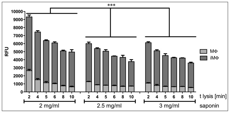
Figure 2. Release of parasites from infected macrophages in 96-well plates.
Bone marrow derived macrophages were infected overnight with L. donovani amastigotes. Cells were incubated for a further 3 days and then treated with DMEM containing the indicated concentration of saponin. Supplemented RPMI was added to the released amastigotes and plates were further incubated for 4 days at 26°C before addition of Alamar blue and subsequent measurement of fluorescence in a plate reader. The survival of freed amastigotes is dependent on the concentration of saponin used and the time of lysis. A 5-fold greater signal of reduced Alamar blue from freed amastigotes over uninfected macrophages was achieved with 2 mg·ml-1 saponin for 5 min; this concentration was used for all other experiments performed. All samples were run in triplicate; one representative experiment is shown. MΦ, uninfected macrophages; iMΦ, infected macrophages. *** denotes P<0.0001 as determined by a two-way ANOVA.

