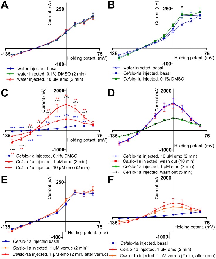
Figure 5. Determination of emodepside effects on current voltage curves in Xenopus laevis oocytes injected with Celslo-1a cRNA.
A) Current voltage curves (IVCs) (means ± SEM) were recorded in water-injected oocytes without addition of drugs (basal), after addition of the vehicle (0.1% DMSO, 0.003% Pluronic F-68) and after addition of 10 µM emodepside (emo) (n = 10). B) Currents obtained from oocytes injected with water (n = 10) or Celslo-1a cRNA in the absence of any drug (basal, n = 8) or in the presence of vehicle (0.1% DMSO, 0.003% Pluronic F-68, n = 8). The asterisk highlights a significant difference of both curves obtained from Celslo-1a cRNA injected groups of oocytes to the water-injected oocytes. C) Currents were recorded in Celslo-1a injected oocytes at the presence of vehicle (0.1% DMSO, 0.003% Pluronic F-68), or emodepside (1 µM or 10 µM emo) (n = 10). For 10 µM emodepside the upper asterisks symbolize the comparison to 1 µM emodepside and the lower the comparison to the vehicle control. The asterisks at the 1 µM emodepside curve indicate significant differences to the control. D) Oocytes injected with Celslo-1a were incubated with 1 µM (n = 6) or 10 µM (n = 7) emodepside (emo) before recording the IVC curves. Then, oocytes were perfused with normal frog ringer for 5 or 10 min, respectively, before a second IVC was recorded from the same oocytes (n = 7). E) Oocytes were preincubated in the absence of drugs (basal) or with 1 µM verruculogen (verruc) before IVCs were recorded. Then, oocytes were perfused for 2 min before 1 µM emodepside (emo) was added (n = 8). F) Oocytes injected with water (n = 10) or with Celslo-1 cRNA (n = 6) were incubated in the absence of drugs (basal) or with 1 µM emodepside before IVCs were recorded. After perfusion with normal frog ringer for 2 min, verruculogen (verruc) was added for 2 min followed by recording of IVCs.*, p<0.05; **, p<0.01; ***, p<0.001.

