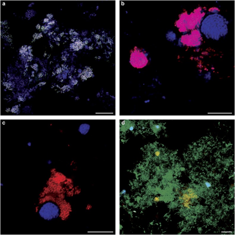
Figure 2.
Confocal micrographs of FISH-stained Nitrotoga-like bacteria in activated sludge samples from WWTPs Bad Zwischenahn (a–c) and Deuz (d). (a) Nitrotoga cell aggregates hybridized to probes Ntoga122 (green), FGall221b (red) and EUB338mix (blue). Nitrotoga appears white because of overlay of all probe signals. (b) Nitrotoga detected by probes FGall221b (red) and EUB338mix (blue) at high magnification. Nitrotoga appears magenta. (c) Simultaneous detection of Nitrotoga and AOB cell clusters by probes FGall221b (red) and Cluster6a192 (blue) at high magnification. Note the close vicinity of Nitrotoga and AOB, reflecting their metabolic interaction. (d) Simultaneous detection of Nitrotoga and Nitrospira by probes Ntoga122 (red), Ntspa662 (blue) and EUB338mix (green). Nitrotoga appears yellow, and Nitrospira cyan. For probe details refer to Table 2 and Supplementary Table S1. The scale bar in all micrographs=10 μm.

