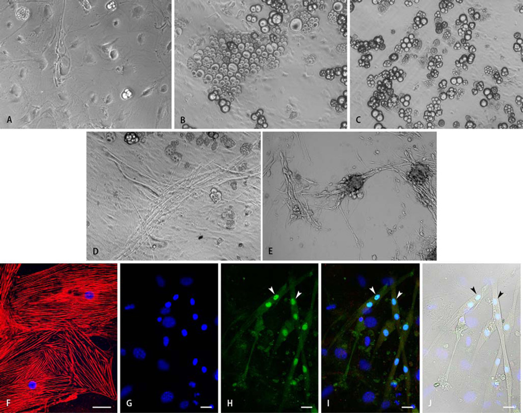
Fig. 2.
Representative light microscopic images of SM+ cells after 30 days of differentiation demonstrating the effects of culture media composition on cellular morphology. Panels a–c show the heterogeneous (epithelial, endothelial, adipose, and others) morphology of SM+ cells cultured in basic medium (BM) containing IGF-1 and dynorphin B (a), and BM supplemented with insulin (b), or oxytocin (c). Exposure of SM+ cells to insulin or oxytocin increased differentiation into an adipose phenotype. Supplementation of BM with bFGF (d) or TGF-β1 (e) resulted in enhanced myotube formation from SM+ cells. Morphological observations were confirmed via immunostaining (f–j). Panel f shows differentiation of SM+ cells cultured in BM supplemented with insulin into epithelial cells positive for the epithelial cytoskeletal marker cytokeratin 17 (red). Panels g–j show representative images of SM+ cells stained for the cardiac-specific marker cardiac myosin heavy chain (i,j red), and skeletal muscle-specific transcription factor myogenin (h–j, green). Nuclei are stained with DAPI (f,g,i,j, blue). Myogenin expression was localized to nuclei (H,l, exemplified by arrowheads). Nuclei expressing myogenin (h–j, exemplified by arrowheads) belonged to cells negative for cardiac marker in red fluorescence (h,j) and exhibited morphology characteristic of myotubes (j). Scale bar = 40 µm (f) or 20 µm (g–j)

