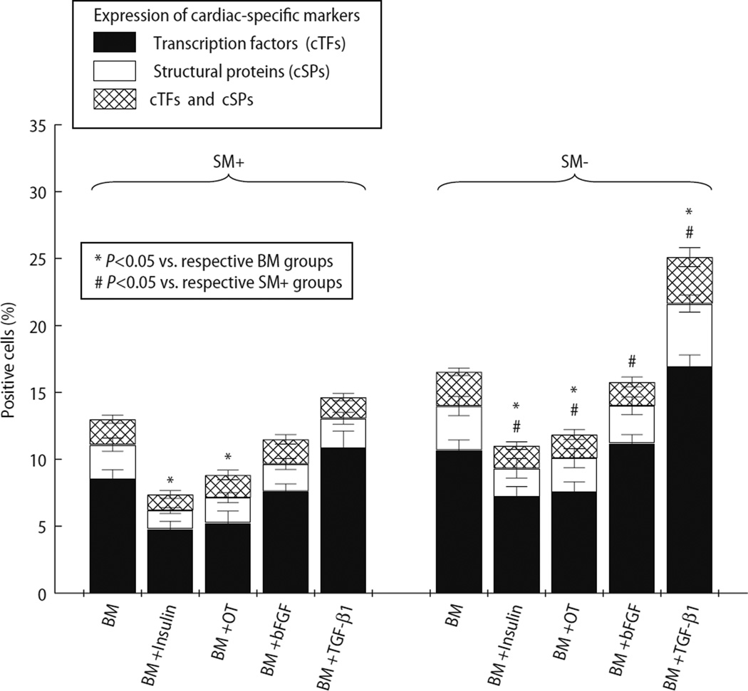
Fig. 5.
Percentage of cells positive for cardiac-specific markers (Y axis), demonstrating the effects of different media (X axis) on cardiomyogenic differentiation of SM+ and SM− cells. Cells positive for cardiac transcription factors (cTFs) and/or structural proteins (cSPs) were counted and expressed as a percentage of total cells. The percentage of cells positive for cTFs is represented by solid bars, cSPs by white bars, and cells positive for both by cross-hatched bars. Cardiomyocytic differentiation was noted in both SM+ and SM− populations. However, compared with SM+ cells, SM− cells exhibited greater cardiomyogenic potential in all media. Supplementation of BM with TGF-β1 resulted in cardiomyocytic differentiation in the greatest fraction of cells in both groups

