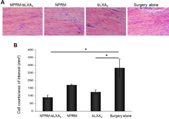
Figure 3.
NPRM-bLXA4 and bLXA4 alone limit inflammatory cell infiltrate. (A) Histological images captured from hemotoxylin and eosin–stained slides at 400× magnification at the subepithelial connective tissue at 3 mo. (B) Inflammatory cell counts were determined using Image J. (*P < 0.05, analysis of variance; n = 4/group). Three captures were performed for each site, and the averages were used for analysis.

