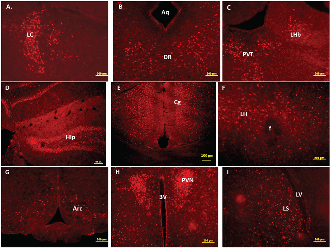
Figure 4.
Immunofluorescence staining in the mouse brain demonstrating representative c-Fos activated regions following intraperitoneal injection of the peripherally-acting nicotine analog, nicotine pyrrolidine methiodide (NIC-PM, 30 µg/kg). A: Locus coeruleus (LC). B: dorsal raphe nucleus (DRN). C: Paraventricular thalamic nucleus (PVT) and lateral habenular nucleus (LHb). D: Hippocampus (Hip). E: Cingulate cortex (Cg). F: Lateral hypothalamus (LH). G: Arcuate hypothalamic nucleus (Arc). H: Paraventricular hypothalamic nucleus (PVN). I: Lateral septal nucleus (LS). J: Medial orbital cortex (MO). K: Piriform cortex (Pir). L: Angular insular cortex (AI) and olfactory nucleus (ON). The data obtained for the NIC treatment at the above representative sites were qualitatively identical to those obtained for the NIC-PM treatment.


