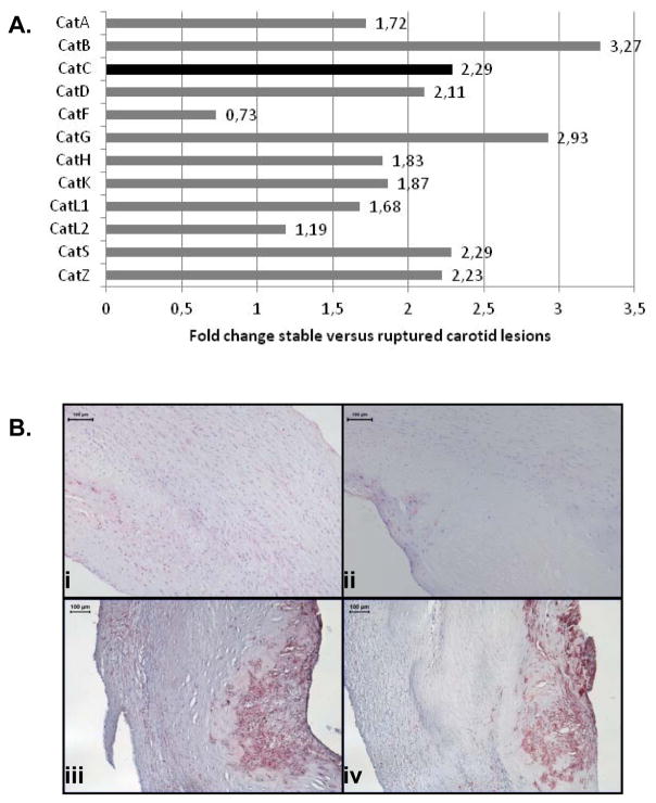
Figure 1.
A: Cathepsin family members were differently expressed in human ruptured carotid endarterectomies, as determined by microarray analysis. Fold changes stable versus ruptured lesion, all p<0.0001. B: Immunohistochemical staining of human carotid atherosclerotic lesions using CatC and CD68 antibodies. Panel (i): representing CatC expression in an early lesion, panel (ii): CatC expression in a stable lesion, panel (iii): CatC expression in a ruptured lesion, panel (iv): parallel section of panel (iii) showing that CatC expression localizes to the same areas of intense CD68-positive staining cells.

