Figure 2.
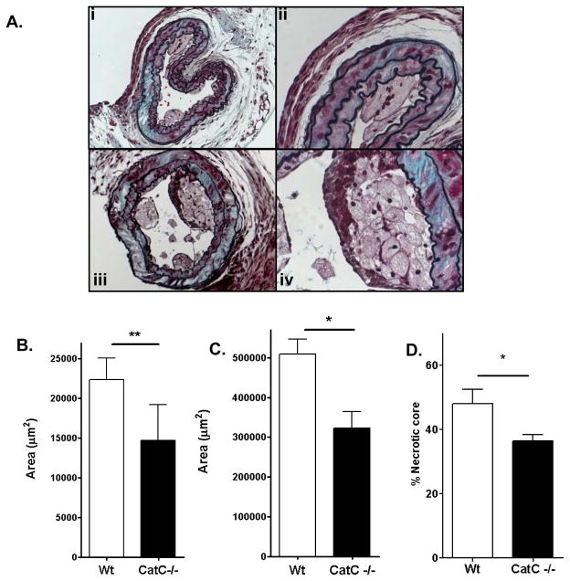
Plaque analysis of mice lesions. A : Movat’s staining of CatC−/− (i) and WT (iv) lesions. Panel (ii) shows the presence of multinucleated cells in a lesion from a CatC−/− chimeric mouse, panel (iii) the presence of a fibrous cap in a lesion from a WT mouse. Further analysis is shown in B: plaque area of collar-induced atherosclerotic lesions in the carotid artery, C: plaque area of advanced lesions in the aortic root. D:necrotic core area of advanced lesions in the aortic root * = p<0.05, ** p=<0.01.

