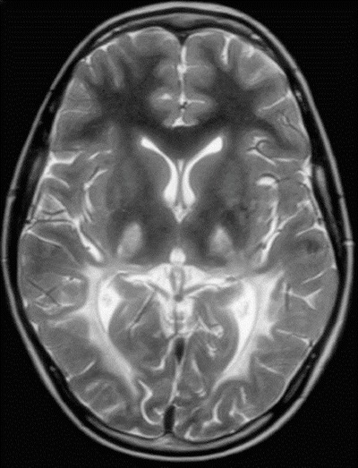
Fig. 1.

MRI brain scan at 10 years of age. A representative image of the T2-weighted sequence is shown demonstrating changes in signal intensity within the posterior white matter in a bilateral, symmetric fashion as well as increased signal intensity within the thalami
