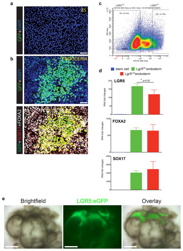
Extended Data Figure 7. Characterization of LGR5: eGFP BAC transgenic reporter hESC line.
a, H9-LGR5:eGFP hESC line did not show eGFP fluorescence in undifferentiated, pluripotent stem cells. b, Upon differentiation to definitive endoderm, robust eGFP expression was observed, consistent with published microarray and RNA-sequencing analyses that show LGR5 as a highly enriched endoderm transcript6,30. The top panel shows DAPI and eGFP staining, whereas the bottom panel shows eGFP co-localization with endoderm markers SOX17 and FOXA2. c, FACS was used to sort LGR5:eGFPLO and LGR5:eGFPHI from 3-day Activin A-treated definitive endoderm cultures. d, qPCR was used to measure LGR5, FOXA2, and SOX17 expression levels in undifferentiated H9-LGR5:eGFP cells (blue bars; “stem cell”) and in FACS-purified H9-LGR5:eGFP endoderm (red bars, LGR5:eGFPLO; green bars, LGR5:eGFPHI). As expected, LGR5, FOXA2, and SOX17 were all highly enriched in both LGR5:eGFPLO and LGR5:eGFPHI endoderm populations compared to undifferentiated controls, and the LGR5:eGFPHI cells showed significant enrichment of LGR5 mRNA, but not FOXA2 or SOX17, compared to the LGR5:eGFPLo population. n=3 biological replicates for each group and error bars represent the S.E.M. *, p<0.05. This analysis suggests that the LGR5:eGFP BAC construct drives eGFP expression in endoderm cells with the highest levels of LGR5 expression. e, H9-LGR5:eGFP hESCs were differentiated into antral gastric organoids. Bright field and GFP stereomicrographs of 30-day hGOs showed that the organoid epithelium developed regionally-restricted areas of LGR5:eGFP expression, suggesting that LGR5+ stem cell populations formed during the differentiation of the organoids. Scale bars, 100 μm.

