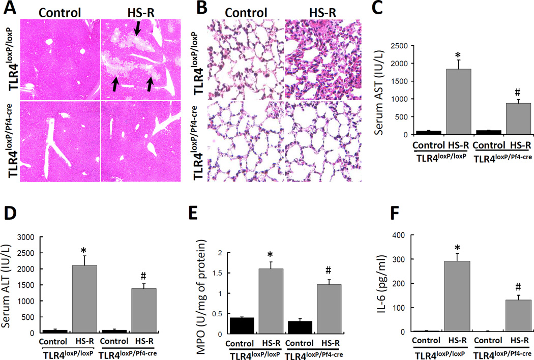
Figure 7.
Effect of HS-R on lung and liver injury and cytokine release. TLR4loxP/Pf4-cre mice and TLR4loxP/loxP mice were subjected to HS-R or unmanipulated control and liver and lung injury was assessed by histology, circulating AST and ALT concentrations, lung MPO activity and circulating IL-6. Liver (A) and lung (B) were sectioned and stained with H&E and tissue damage induced by HS-R was assessed by light microscopy (Nikon FX series). Black arrows show the regions of necrosis. (Original magnification ×100). Serum AST (C) and ALT (D) levels were assayed and are presented as means ± SEM (n = 8–10 mice/group). Aliquots of the mouse lung tissues were homogenized to prepare total protein samples and MPO levels were assayed by ELISA (E). The levels are presented as means ± SEM (n = 8–10 mice/group). Serum samples from mice treated as described above were used to assay IL-6 concentration by ELISA (F) and the levels are presented as means ± SEM (n = 8–10 mice/group). *p<0.05 vs. control. #p<0.05 vs. HS-R group of TLR4loxP/loxP mice.

