
Fig. 3. Effect of anti-CIRP antibody treatment on tissue damage and cellular architecture in the liver after hepatic I/R.
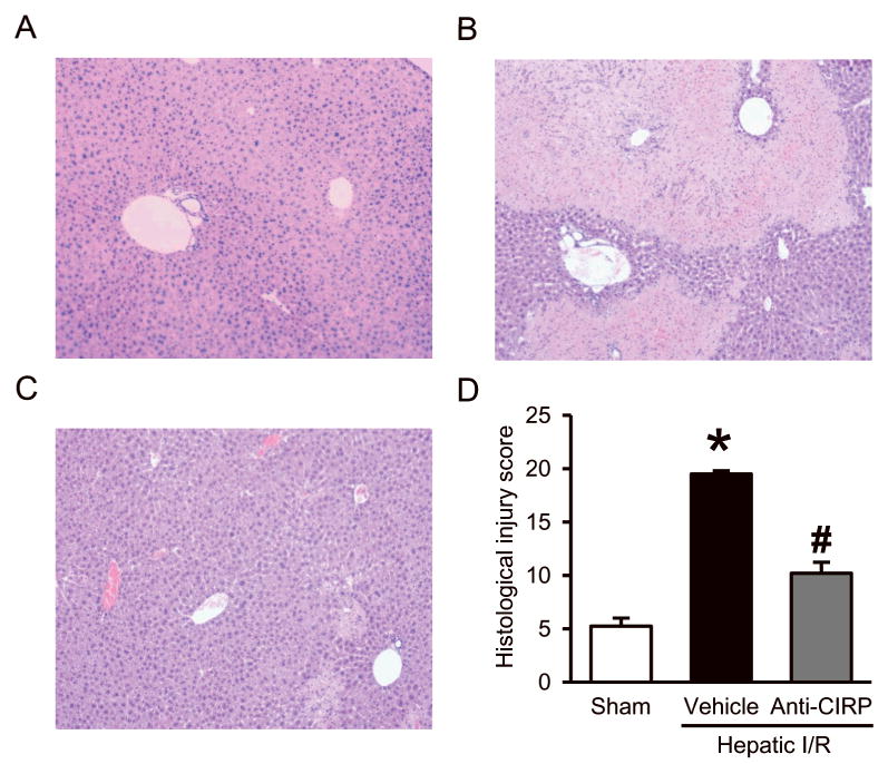
Histologic evaluation of the liver in the sham (A), vehicle (B), and anti-CIRP antibody-treated (C, Anti-CIRP) groups. Liver tissues were harvested 24 h after hepatic I/R, processed, and then stained with hematoxylin-eosin (H&E). Representative photomicrographs at 100× magnification. D, Semiquantitative histologic injury score measuring differences in cytoplasmic vacuolization, cytoplasmic fading, nuclear condensation, nuclear fading, nuclear fragmentation, and erythrocyte stasis examined on H&E staining as described in Materials and Methods. Data presented as means ± SE (n=4-5/group) and compared by one-way ANOVA and SNK method; *p < 0.05 vs. sham; #p < 0.05 vs. vehicle.