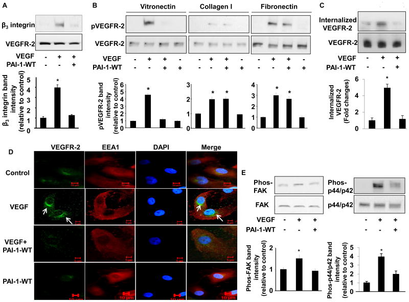
Figure 1.
PAI-1 inhibits VEGFR-2 signaling. (A) PAI-1 inhibits β3 integrin-VEGFR-2 complex formation. HUVECs were cultured on VN in presence of vehicle control, VEGF (50 ng/mL), or VEGF and PAI-1-WT (10 μg/mL), as indicated (“+” = added, “-” = omitted, replaced by vehicle). Cell lysates were prepared and incubated with resin-bound anti-VEGFR-2 antibody. Captured proteins were analyzed by Western blotting with anti-β3integrin antibodies. Representative images of 3 independent experiments are shown. *P<0.05 vs. negative control. (B) Inhibition of VEGF-induced VEGFR-2 phosphorylation by PAI-1 is VN-dependent. HUVECs were cultured on VN, collagen, or fibronectin in presence of vehicle control, VEGF (50 ng/mL), VEGF and PAI-1-WT (10 μg/mL), or PAI-1-WT, as indicated. Cell lysates were prepared and subjected to Western blotting to detect phosphorylated (p) and total VEGFR-2. (C) PAI-1 inhibits VEGFR-2 endocytosis. HUVEC cell surface proteins were biotinylated, after which cells were cultured in presence of vehicle control, VEGF, or VEGF and PAI-1-WT, as shown. Internalized VEGFR-2 was detected by Western blotting. (D) PAI-1 inhibits translocation of VEGFR-2 to perinuclear endosomes. HUVECs were cultured on VN in presence of vehicle control, VEGF (50 ng/mL), VEGF and PAI-1-WT (10 μg/mL), or PAI-1-WT, as indicated. VEGFR-2 and EEA1 were detected by immunofluorescence confocal microscopy. DAPI (nuclear) and merged VEGFR-2/EEA1/DAPI images are also shown. Images are representative of 3 independent experiments. Arrows indicate perinuclear location of VEGFR-2. (E) PAI-1 inhibits intracellular signaling pathways downstream of VEGFR-2. HUVECs were cultured on VN in presence of vehicle control, VEGF, or VEGF and PAI-1-WT, as indicated. Cell lysates were prepared and subjected to Western blotting to detect phosphorylated and total FAK and p44/p42. All graphs correspond to blots above them and represent densitometric analyses of 3 independent experiments. *P<0.05 vs. negative control group (1st bar).

