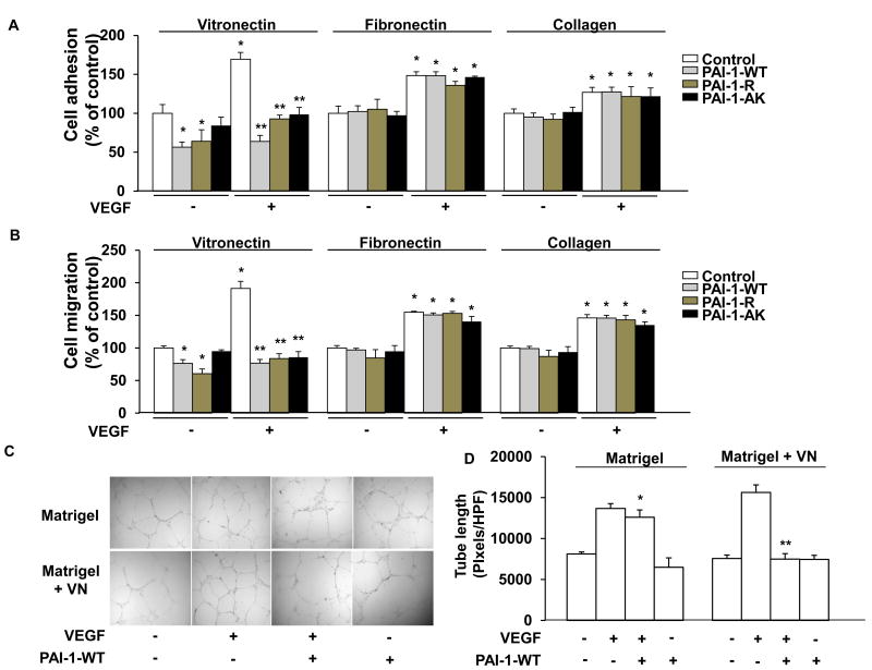
Figure 3.
PAI-1 inhibits VEGF-induced HUVEC adhesion, migration, and tubule formation in a VN-dependent manner. (A,B) HUVECs were added to wells coated with VN, fibronectin, or collagen, as shown, incubated in the presence of recombinant PAI-1 (WT, R, or AK forms, 10 μg/mL) or vehicle control, as shown, and treated with VEGF (+, 50 ng/mL) or vehicle control (-), as indicated, after which cell adhesion (A) and migration (B) were measured. Data are mean of triplicate experiments. *P<0.05 vs. negative control (HUVECs not exposed to PAI-1 or VEGF). **P<0.05 vs. HUVECs incubated with VEGF only (no PAI-1). (C) Tubule formation. Representative images of HUVECs grown on Matrigel or Matrigel supplemented with VN (10 μg/mL) in the absence (-) of added VEGF or PAI-1-WT or in the presence (+) of VEGF (50 ng/mL), VEGF and PAI-1-WT (10 μg/mL), or PAI-1-WT, as shown. (D) Quantitative assessment of quadruplicate tubule formation experiments. *P>0.05 vs. HUVECs grown on Matrigel and exposed only to VEGF. **P<0.05 vs. HUVECs grown on Matrigel containing VN and exposed only to VEGF.

