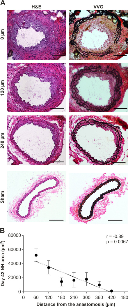
Figure 1.
Representative AVF pathology at day 42 after AVF creation. A. Neointimal hyperplasia was evident in the inflow arterial limb of the AVF (hematoxylin and eosin (H&E), left column; Van Gieson’s stain (VVG), right column; Scale bar, 100 µm). B. Correlation between the neointimal area in day 42 AVF as a function of increasing distance from the anastomosis (r=−0.89, p=0.0067, n=7 mice). Error bar, S.E.M.

