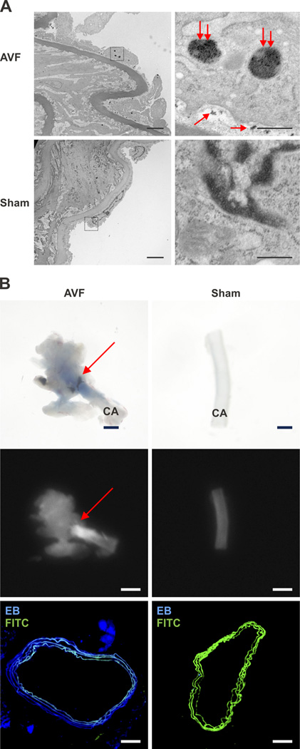
Figure 3.
Assessment of CLIO-VT680 nanoparticle localization within the arterial segment of AVF. A. Transmission electron micrographs of a representative day 14 CLIO-VT680 injected animal. In AVF, CLIO-VT680 nanoparticles (double red arrows, large nanoparticle clusters) were found inside the endothelial cells and between the endothelial cells and the basement membrane (single red arrows, smaller nanoparticle collections). There was minimal CLIO-VT680 particles deposition in sham-operated control arteries (lower row). Left column panel: scale bar, 4 µm. Right column panel: scale bar, 0.5 µm. IEL, internal elastic lamina. B. Light microscopy (top row) and fluorescence reflectance imaging (middle column) of day 14 AVF showed Evans blue deposition in the juxta-anastomotic AVF zone, but only minimally in the sham-operated contralateral carotid artery (scale bar: 1 mm). Fluorescence microscopy (lower row) showed strong Evans blue signal (blue) in the AVF carotid arterial wall, with little signal in sham-operated contralateral arteries. Scale bar, 100 µm. CA, carotid artery, EB, Evans Blue, FITC, fluorescein isothiocyanate.

