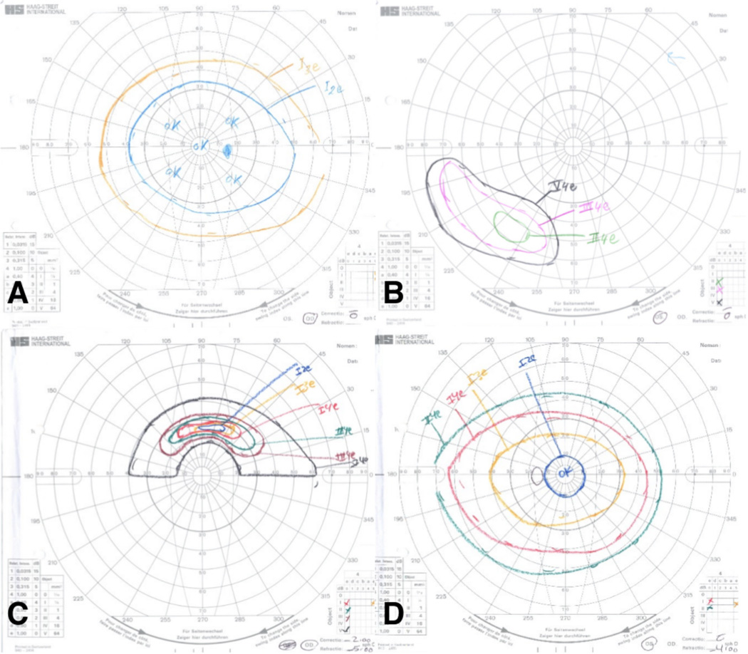
Figure 2. Goldmann visual field examples): (A,B: patient 9.
A: The preferred right eye has a slightly constricted field that is otherwise normal; B: The non-preferred left eye has only an inferotemporal island of vision. (C,D; patient 16): C: The non-preferred right eye has only a superior arcuate island of vision; D: The preferred left eye has a slightly constricted field that is otherwise normal.

