
Figure 1. Photos of CT and gross specimen.
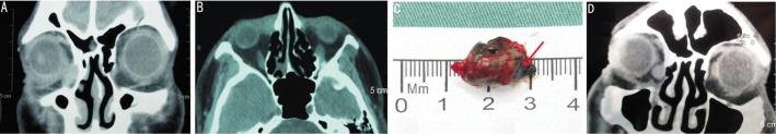
A: Photo of the patient showed edema of the right medial canthal area; B: Coronal CT scans showed a soft tissue mass in the right lacrimal sac (arrow); C: En bloc resection of the melanoma, whose transverse section was dark (arrow); D: CT showed local recurrence of melanoma one year later (arrow).