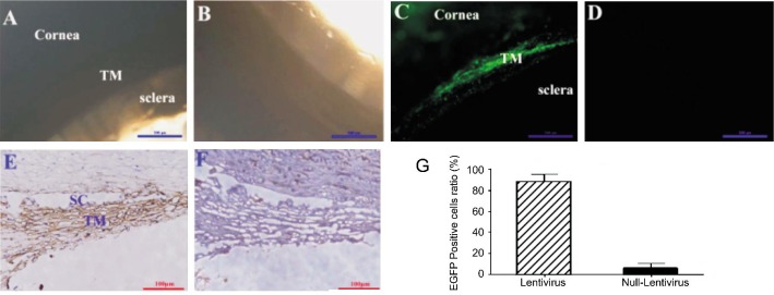
Figure 3. Genetic modification of HTM cells in an ex vivo model of cultured anterior segment of human eye.
Cultured anterior segments of the human eye were transfected with HIV-based lentivirus which was delivered by perfusion system. Twenty-one days later, EGFP expression could be detected by fluorescence microscopy in transfected cells (A: Brightfield image, C: Fluorescent image, scale bar=500 µm), but not in control group (B: Brightfield image, D: Fluorescent image; scale bar=500 µm). Further immunochemistry study showed that EGFP was expressed both in TM and Schlemm's canal (E for EGFP contained HIV-based lentivirus, F for control virus, scale bar=100 µm). EGFP positive cell ratio were calculated by counting positive cells and the total cell nucleus (G for positive cell ratio, t-test, P<0.01).

