
Figure 2.
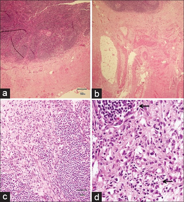
Histopathology specimen from lymph nodes and omentum showed extensive perinodal fibrosis (a, b) with the presence of focal dense lymphoplasmacytic infiltrate (arrows) and loose fibrovascular proliferation infiltrating adipose tissue (c, d)
Official websites use .gov
A
.gov website belongs to an official
government organization in the United States.
Secure .gov websites use HTTPS
A lock (
) or https:// means you've safely
connected to the .gov website. Share sensitive
information only on official, secure websites.
Histopathology specimen from lymph nodes and omentum showed extensive perinodal fibrosis (a, b) with the presence of focal dense lymphoplasmacytic infiltrate (arrows) and loose fibrovascular proliferation infiltrating adipose tissue (c, d)