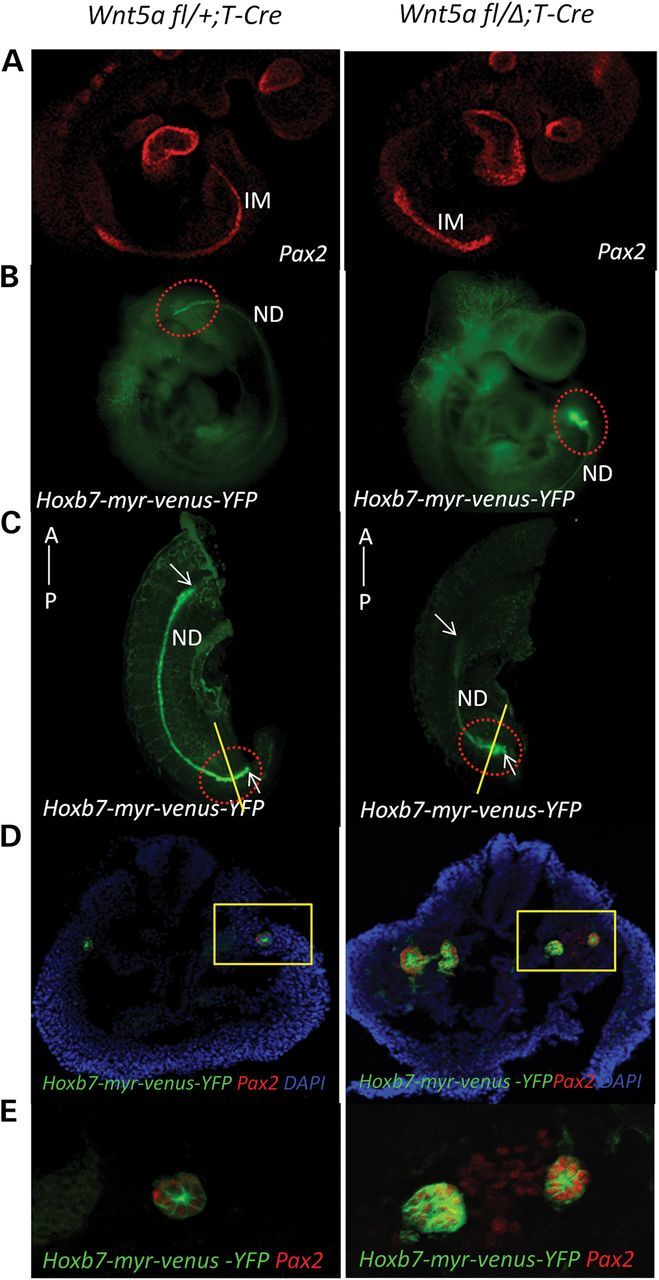
Figure 4.

The IM is shortened and broadened in the E9.5 Wnt5a mutant and manifests a duplicated ND at its caudal terminus. (A) Embryos at E9.5 were stained with Pax-2 antibody. left: Wnt5a fl/+;T-Cre; right: Wnt5a fl/Δ;T-Cre. (B) left: Wnt5a fl/+;T-Cre;Hoxb7-myr-venus-YFP embryo at E9.5; right: Wnt5a fl/Δ;T-Cre;Hoxb7-myr-venus-YFP embryo at E9.5. A red-dotted ellipse marks the posterior ends of the embryos. (C) Embryo images without the head using light sheet microscopy. A red-dotted ellipse marks the posterior end of the embryos. White arrows mark the anterior and posterior ends of the ND. (D) Transverse sections of the posterior end of the IM in E9.5 embryos were stained for Pax-2 expression. left: Wnt5a fl/+;T-Cre;Hoxb7-myr-venus-YFP embryo at E9.5; right: Wnt5a fl/Δ;T-Cre;Hoxb7-myr-venus-YFP embryo at E9.5. A yellow rectangle marks the IM. (E) Enlarged image of the IM in (D).
