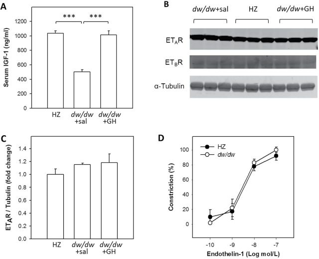
Figure 1.
Validation of the insulin-like growth factor (IGF)-1 deficiency in the Lewis dwarf rats and characterization of the vascular responses to endothelin-1. (A) Serum IGF-1 levels were determined by enzyme-linked immunosorbent assay. One-way analysis of variance (ANOVA) revealed a significant difference among groups (p < .001) and pairwise comparisons demonstrate that the serum IGF-1 level in the dw/dw+sal group is significantly reduced compared to the HZ group (p < .001) and growth hormone–treated group (p < .001). Homogenates of cerebral cortex from a separate cohort was used for Western blotting to semiquantify the expression of A-type (ETAR) and B-type (ETBR) receptors. Representative images are shown in (B) and quantitation of ETAR expression is shown in (C). One-way ANOVA demonstrated that ETAR expression is similar among three groups (p = .389). Expression of ETBR was too low to be quantitated but appeared not to be different among groups. n = 3/group. (D) Constriction of cannulated middle cerebral arteries isolated from dw/dw and HZ rats in response to increasing concentrations of endothelin-1. Constriction was calculated as a percentage of changes in diameter from the baseline and normalized to the maximum constriction which was reached at 100nM. There is no significant difference between the endothelin-1 -induced constriction of middle cerebral arteries isolated from dw/dw and HZ rats. Data are mean ± SEM (n = 5/group).

