FIG. 3.
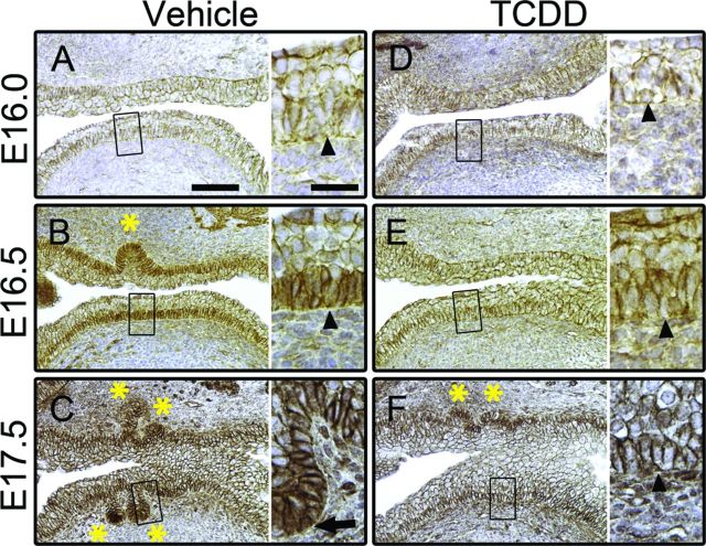
TCDD prevents accumulation and nuclear localization of NP-CTNNB1. C57BL/6J mouse embryos were exposed in utero to vehicle or TCDD (5 μg/kg dam, po) at E15.5 and UGSs were harvested at E16.0, E16.5, or E17.5. UGSs were sectioned along the sagittal plane and the anterior and ventral budding regions are shown (bladder is to the right in each image). Images are representative of at least three litter-independent UGSs per treatment group. In each panel, the image on the left is shown at 100X (scale bar represents 100 μm) and the boxed region is shown on the right at 200X (scale bar represents 25 μm). IHC staining using DAB as the chromogen (brown) shows subcellular localization of NP-CTNNB1; nuclei are counterstained with hematoxylin (blue). Arrowheads point to ventral basal epithelial cells; asterisks mark prostatic buds. In E16.0 vehicle-exposed UGSs, NP-CTNNB1 is restricted to cell membranes (A). By E16.5 in vehicle-exposed UGSs cytoplasmic accumulation and nuclear localization of NP-CTNNB1 is detectable in the ventral basal epithelial cells—note that the nuclei of intermediate epithelial cells are devoid of CTNNB1 staining (B). At E17.5, in vehicle-exposed UGSs, anterior and ventral prostatic buds are visible, with NP-CTNNB1 nuclear localization primarily seen in bud tips (arrow in C). In utero exposure to TCDD has no effect on subcellular localization of NP-CTNNB1 at E16.0 (D). However, at E16.5 (E) and E17.5 (F) TCDD-exposed UGSs show no NP-CTNNB1 accumulation in the cytoplasm, no NP-CTNNB1 nuclear localization, and no ventral buds.

