
FIGURE 1.
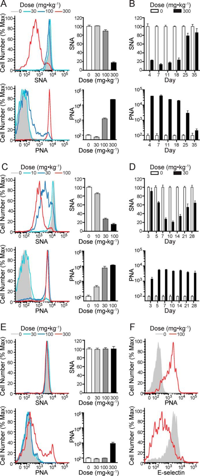
Administration of 3F-NeuAc to mice results in dose- and time-dependent decreases in sialoside expression on peripheral blood leukocytes. A, SNA (upper panels) and PNA (lower panels) staining on B cells 11 days after mice received one of the indicated single intravenous doses of 3F-NeuAc. B, time course summary of SNA (upper panels) and PNA (lower panels) staining on B cells from mice that received a single 300 mg·kg−1 intravenous injection of 3F-NeuAc. C, SNA (upper) and PNA (lower) staining on B cells (day 11) from mice that received a daily intraperitoneal injection of 3F-NeuAc at one of the indicated doses for 7 days. D, time course summary of SNA (upper) and PNA (lower) staining on B cells from mice that received a daily 30 mg·kg−1 intraperitoneal injection of 3F-NeuAc for 7 days. E, SNA (upper) and PNA (lower) staining on T cells 11 days after mice received one of the indicated single intravenous doses of 3F-NeuAc. F, PNA (upper) and E-selectin (lower) staining on granulocytes (day 7) from mice that received a daily intraperitoneal injection of 100 mg·kg−1·day−1 3F-NeuAc for 7 days. All data represent the means ± S.E. of four replicates, are expressed as percentages relative to mice that received vehicle, and are representative of three independent experiments. Max, maximum.