FIGURE 8.
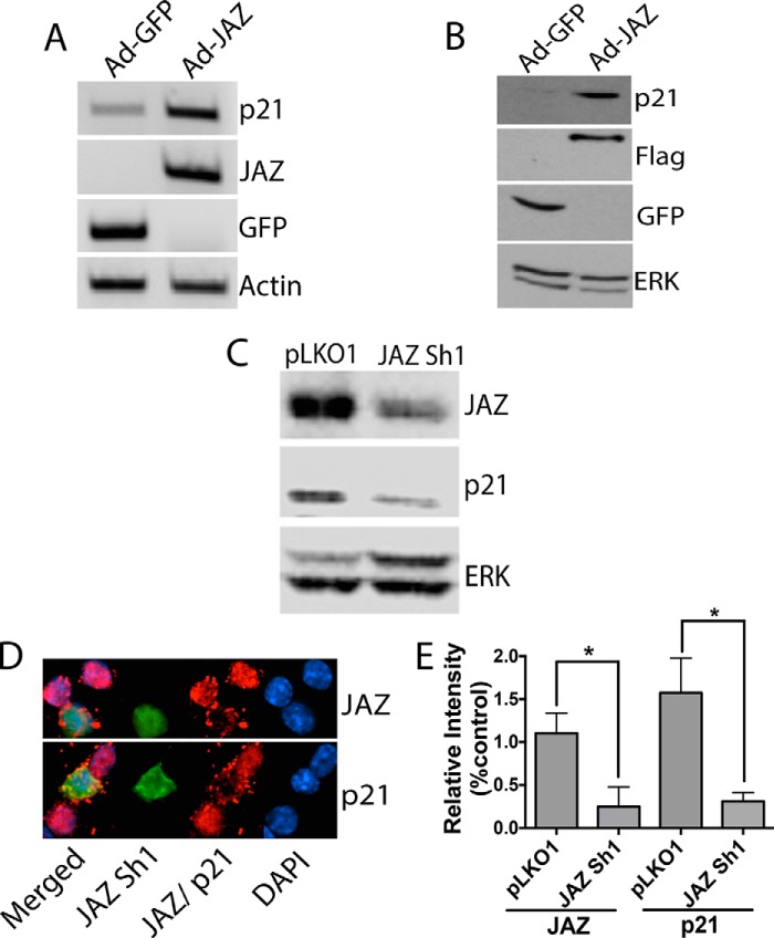
JAZ stimulates p21 expression. A, CGNs were infected with Ad-GFP or Ad-JAZ-FLAG for 36 h after which RNA was prepared and RT-PCR analysis was performed using primers for p21, GFP, and JAZ. Actin serves as a normalization control. B, cell lysates were prepared from CGNs infected with Ad-GFP or Ad-JAZ-FLAG for 36 h. Western blot analysis was performed using a p21 antibody. GFP and FLAG antibodies were used to evaluate infection efficiency of both GFP and JAZ adenovirus. ERK was used as a loading control. C, HT22 cell lines were transfected with either pLKO1 or JAZ Sh1 for 72 h. Cell lysates were collected, and Western blot analysis was performed using JAZ and p21 antibodies. ERK was used as a loading control. D, CGNs were co-transfected with pLKO1 (control) and JAZ Sh1 along with GFP (6.5:1) for 48 h and then switched to HK for 24 h. Immunocytochemistry was performed with JAZ (top) and p21 (bottom) antibodies. GFP fluorescence was used to visualize shRNA-transfected cells. E, quantification of JAZ and p21 immunoreactivity in cells overexpressing JAZ Sh1 or pLKO1 (control). Left asterisk: *, p < 0.05 JAZ immunoreactivity in JAZ Sh1-transfected cells as compared with pLKO1-transfected cells. Right asterisk: *, p < 0.05, p21 immunoreactivity in JAZ Sh1-transfected cells as compared with pLKO1-transfected cells.

