FIGURE 1.
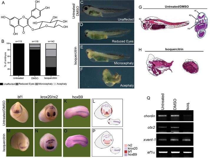
Isoquercitrin induces axial defects in Xenopus embryos. A, molecular structure of isoquercitrin. B, quantification of the phenotypes obtained by isoquercitrin at 150 μm treatment and control (DMSO). C, control embryo. D–F, isoquercitrin leads to reduced eyes (D), microcephalic (E), or acephalic (F) phenotypes. G and H, histological sections show lack of dorsal structures after isoquercitrin treatment. In situ hybridization reveals disturbance in expression domain of bf1 (I and M), krox20/rx2 (J and N), and hoxB9 (K and O). L and P, schematic representation of the alterations in the gene expression of anterior region of the embryo. Q, PCR reveals reduced expression of anterior markers chordin and otx2, whereas xvent-1 is unaffected after isoquercitrin (Isoq.) treatment (lane 3). Ef1-α was used as loading control.

