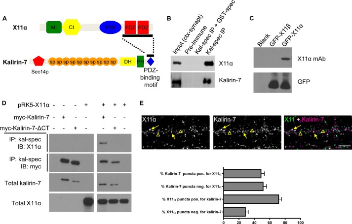
FIGURE 1.
Kalirin-7 and X11α interact and co-localize in dendrites. A, structures of X11α and kalirin-7. Bar along kalirin-7's C terminus indicates the region used as bait in a yeast two-hybrid screen; bar along X11α's sequence indicates sequence contained in a clone that interacted with kalirin-7 bait (10). MI, Munc-18-interacting region; CI, CASK-interacting region; PTB, phosphotyrosine-binding domain; PDZ, PSD-95/Discs large/ZO-1 domain; Sec14p, Sec14p-like domain; sp, spectrin-like domain; DH, Dbl-homology domain; PH, pleckstrin homology domain. B, co-immunoprecipitation (IP) of X11α with kalirin-7 from rat cortical synaptosomes (ctx-synapt). X11α is present in the input lane and in the IP lane, but absent when a GST-tagged spectrin region of kalirin-7 interferes with antigen binding. C, X11α-specific antibody detects GFP-tagged X11α, but not X11β, in HEK293 lysates expressing the indicated constructs. D, co-immunoprecipitation of X11α with Myc-tagged kalirin-7 in transiently transfected HEK293 cells is abrogated by truncation of kalirin-7's C-terminal PDZ-binding domain. E, X11α and kalirin-7 co-localize in dendrites of cultured cortical pyramidal neurons. Yellow arrows indicate puncta of co-localization, and yellow arrowheads indicate X11α puncta that do not localize with kalirin-7. Bar graph indicates quantitative measures of respective co-localization of immunofluorescent puncta.

