FIGURE 2.
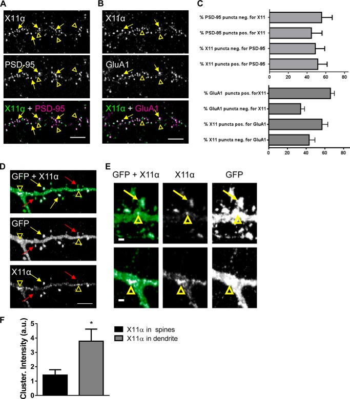
X11α is enriched in cortical synapses and localizes to dendritic spines. A, X11α partially co-localizes with PSD-95 in cultured rat cortical pyramidal neurons (DIV 24). Yellow arrows indicate sites of co-localization, and yellow arrowheads indicate X11α puncta that do not localize with PSD-95 (overlay of green and magenta = white). B, X11α partially co-localizes with AMPA receptor subunit GluA1 in cultured rat cortical pyramidal neurons. Yellow arrows indicate sites of co-localization, and yellow arrowheads indicate X11α puncta that do not localize with GluA1. C, bar graphs are quantitative measures of respective co-localization of immunofluorescent puncta (PSD-95 and X11α; GluA1 and X11α). D and E, representative confocal images of cortical neurons (DIV 24) expressing GFP and pRK5-X11α. Overexpressed pRK5-X11α localizes to dendritic spine heads (yellow arrows) and dendritic shaft (yellow arrowheads). Red arrows indicate dendritic spines containing little or no X11α signal (D). X11α accumulated at the base of spines and at dendritic branching points (yellow arrowheads) (E). F, mean fluorescence intensity of X11α signal in dendritic spines is increased compared with X11α signal in dendritic shaft (*, p < 0.05, Student's unpaired t test). Scale bars, 5 μm (A, B, and D) and 1 μm (E).

