FIGURE 3.
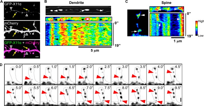
Trafficking of X11α in cortical neurons. A, GFP-tagged X11α localizes to dendrites (white arrowheads) and dendritic spines (yellow arrows) in mCherry-expressing cortical neurons. B, top, image of a segment of dendrite showing mobile (arrows) and stationary (arrowheads) GFP-X11α puncta. Bottom, kymograph (pseudocolored) of GFP-X11α in dendrite segment. Vertical bands of high fluorescence intensity indicate stable fluorescent puncta (arrowheads), although diagonal bands (arrows) indicate mobile puncta. Multiple X11α puncta displayed movement within the dendritic shaft. C, representative kymograph analysis (19 s) of GFP-X11α puncta within the dendritic spine (left). Both mobile (arrows) and stationary (arrowhead) GFP-X11α puncta can be seen in spines. D, time-lapse imaging of GFP-X11α immunofluorescence in dendritic spine (outlined). Red arrows indicate the movement of X11α puncta into and out of a spine during a 10-s imaging session. Scale bars, 5 μm (A), 5 μm (B), and 1 μm (C and D).

