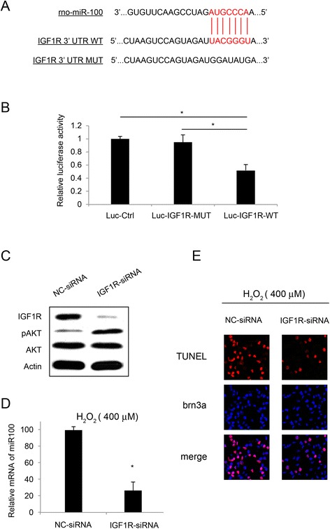
Figure 1.

miR-100 interacted with IGF1R in RGC-5 cells. (A) Schematic diagram was shown for the predicted binding between rno-miR-100 and IGF1R 3’-UTR. The mutated 3’-UTR of IGF1R (IGF1R-mu) was also demonstrated. (B) In a luciferase report assay, HEK 293 T cells were transfected with pmiR-REPORT control vector (Luc-Ctrl), IGF1R vector with mutated 3’-UTR (Luc- IGF1R -mu) or IGF1R with wild-type 3’-UTR (Luc- IGF1R), along with β-galactosidase and miR-100 mimics for 24 hours. Luciferase signals were measured and normalized to the signal of control vector. (*: P <0.05). (C) RGC-5 cells were treated with IGF1R-siRNA or its non-specific siRNA (NC-siRNA), followed by western blotting on AKT/pAKT and IGF1R in 48 hours. RGC-5 cells were pre-treated with IGF1R-siRNA (100 nM) or its non-specific siRNA (NC-siRNA, 100 nM) for 24 hours, followed by H2O2 (400 μM) treatment for another 24 hours. (D) The mRNA expression level of miR-100 was assessed by qRT-PCR (*: P <0.05). (E) RGC-5 apoptosis was assessed by TUNEL staining.
