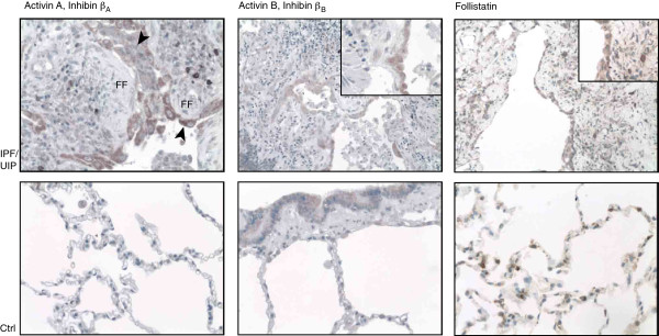
Figure 2.

Activin-A, -B and follistatin immunoreactivity in human usual interstitial pneumonia (UIP) lung tissue and control lung samples. Activin-A immunoreactivity (upper panel, left) is observed in hyperplastic alveolar epithelial cells (arrowheads) overlying fibroblastic foci (FF). Occasionally, macrophage-appearing cells in the lung parenchyma stain positive. Activin-B immunoreactivity is similarly observed in areas of hyperplastic epithelial cells and, to a lesser extent, in parenchymal inflammatory cell infiltrates. Follistatin expression is observed at a high intensity in activated alveolar epithelial cells. Staining is also seen in lung bronchial epithelial cells (activins) and alveolar cells (follistatin), shown in the control (Ctrl) lung sections, lower panel.
