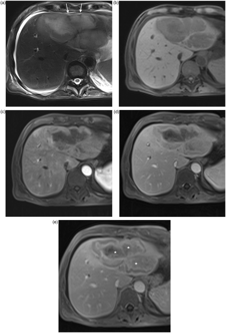
Fig. 13.
A 64-year-old man with inflammatory pseudotumor of liver. Large liver lesion in segments II and IV with capsular retraction (arrows) in high signal intensity on T2W TSE fat-suppressed image (a), low signal intensity on T1W GE image (b). Contrast-enhanced T1W GE images during arterial (c), portal (d), and delayed (e) phases show a fibrous central component with progressive enhancement (*) on portal and late phase although a perilesional halo is enhanced as soon the arterial phase.

