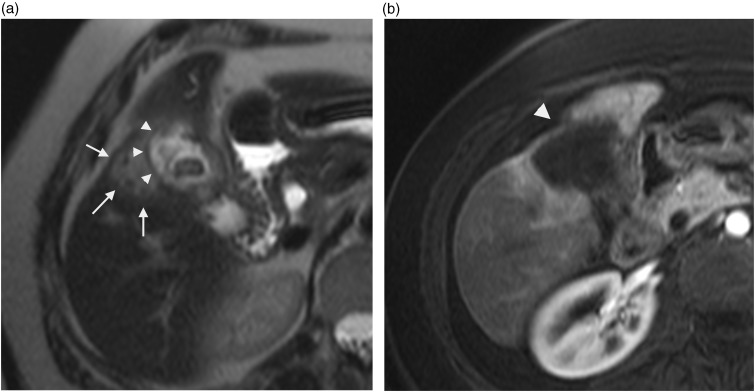
Fig. 5.
A 58-year-old man with gallbladder adenocarcinoma. Axial T2W TSE not fat-supressed MRI (a) shows high intensity infiltrative mass (arrows) with poor-defined interface with gallbladder (arrowheads). Mild progressive peripheral centripetal enhancement was noted on arterial (b), portal and late contrast-enhanced T1W GE images with adjacent hepatic capsular retraction (arrowhead).

