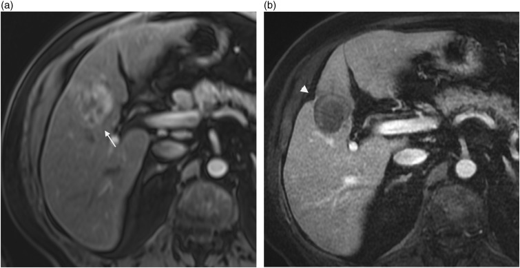
Fig. 9.
A 78-year-old-man with hepatocellular carcinoma. Contrast-enhanced T1W GE (a) image showing an enhanced nodule at the arterial phase in segment 4 prior to treatment (arrow). This lesion was treated with transcatheter chemoembolization and RF ablation; contrast-enhanced T1W GE image after treatment (b) showing associated capsular retraction (arrowhead) and no abnormal enhancement.

