Figure 5.
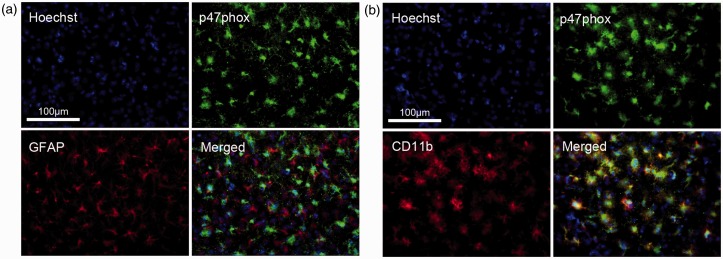
P47phox expression colocalized with microglia, and not astrocytes, in the striatum at 72 h after ischemia/reperfusion.
(a) Fluorescent microscopic image of p47phox (green) and GFAP (red) staining showing no colocalization between the p47phox and astrocytes. (b) Fluorescent microscopic image of p47phox (green) and CD11b (red) staining showing colocalization between the p47phox and microglia; Scale bar = 100 µm in a and b.

