
Figure 1.
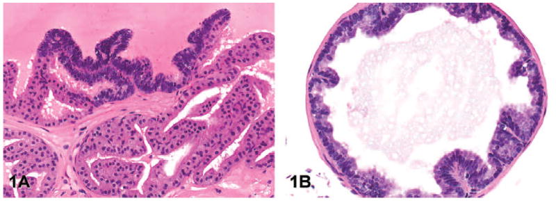
Representative images of low-grade prostatic intraepithelial neoplasia (lesion grade 1) in transgenic adenocarcinoma of the mouse prostate. (A) Note the focal cell stratification, increased nuclear to cytoplasmic ratio, and crowding of the epithelial cells (anterior lobe, hematoxylin and eosin [H&E], magnification 200×). (B) Few, short papillary proliferations of hyperplastic epithelium project into the lumen (ventral lobe, H&E, 200×).