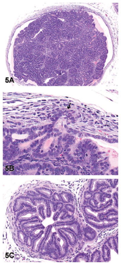
Figure 5.

Representative images of well-differentiated adenocarcinoma (lesion grade 5) in transgenic adenocarcinoma of the mouse prostate. (A) Well-differentiated adenocarcinoma forming a mass of epithelial cells and acini that expands the lumen of a gland from the dorsal lobe of the prostate. Note that this gland is no longer surrounded by a complete or distinct smooth muscle capsule (hematoxylin and eosin [H&E], 40×). (B) Higher magnification of the image in panel A shows invasion of epithelial cells through the basement membrane and into the adjacent stroma (arrow). A stromal reaction in response to invasion is evident as reactive fibroblasts and myoepithelial cells surrounding the invading cells (H&E, 400×). (C) Note the exuberant stromal reaction and replacement of the smooth muscle capsule by reactive fibroblasts in this focus of well-differentiated adenocarcinoma from the lateral prostate (H&E, 200×).
