
FIGURE 1.
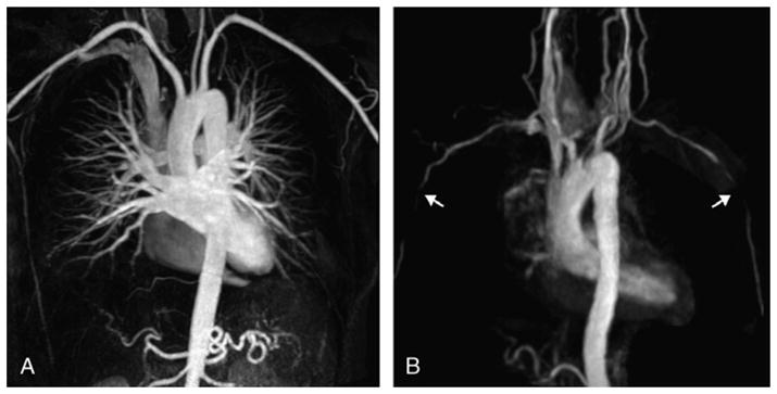
Maximum intensity projection reformations of the aortic arch and supra-aortic branches of a control subject (A) and a patient (B) with biopsy-proven GCA, obtained from contrast-enhanced MRA (fast spoiled 3D gradient echo sequence, breath held, coronal field of view, ECG gated, with 1.0 mmol/kg of gadolinium at a flow rate of 2 mL/s). This patient presented the most found lesions, bilateral stenosis in the axillary arteries (arrows in B). The control subject has normal vessels.