
FIGURE 2.
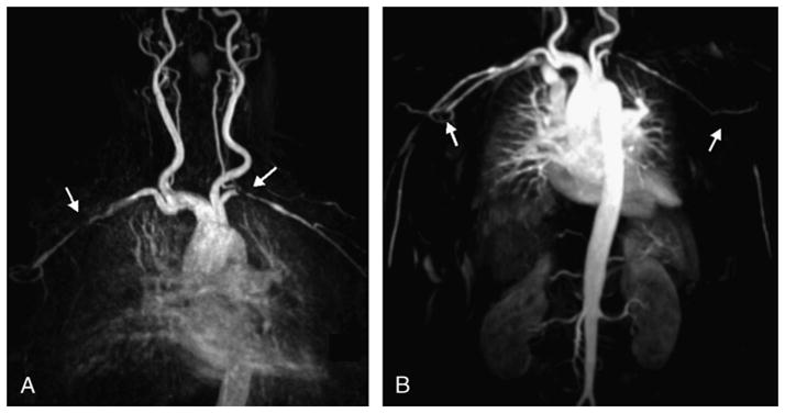
Image (A) in a 70-year-old woman with negative temporal biopsy and symptoms of PMR with aortic arch syndrome and (B) in a 79-year-old woman with biopsy-proven GCA. In both cases, there are lesions in the subclavian and axillary arteries, with bilateral subclavian stenosis in the first patient (arrows in A) and axillary bilateral obstruction in the second patient (arrows in B). Both images are MIP reconstructions from contrast-enhanced MRA (fast spoiled 3D gradient echo sequence, breath held, coronal field of view, ECG gated, with 1.0 mmol/kg of gadolinium at a flow rate of 2 mL/s).