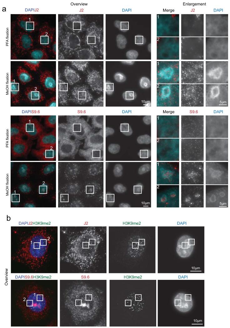
ED Figure 3. Cellular localisation of R-loops, dsRNA and H3K9me2.
a. Immunofluorescence imaging of dsRNA (J2 antibody) and R-loops (S9.6 antibody), using paraformaldehyde (PFA) and methanol (MeOH) as fixing reagents. Fixation with methanol allowed visualisation of R-loops and dsRNA in HeLa cell nuclei. Enlarged boxes (1 and 2) shown in right panels. b. Whole cell images showing immunofluorescence of H3K9me2 with dsRNA (J2-top panel) and R-loops (S9.6-bottom panel). Enlarged versions (1 and 2) are shown in Fig. 2h.

