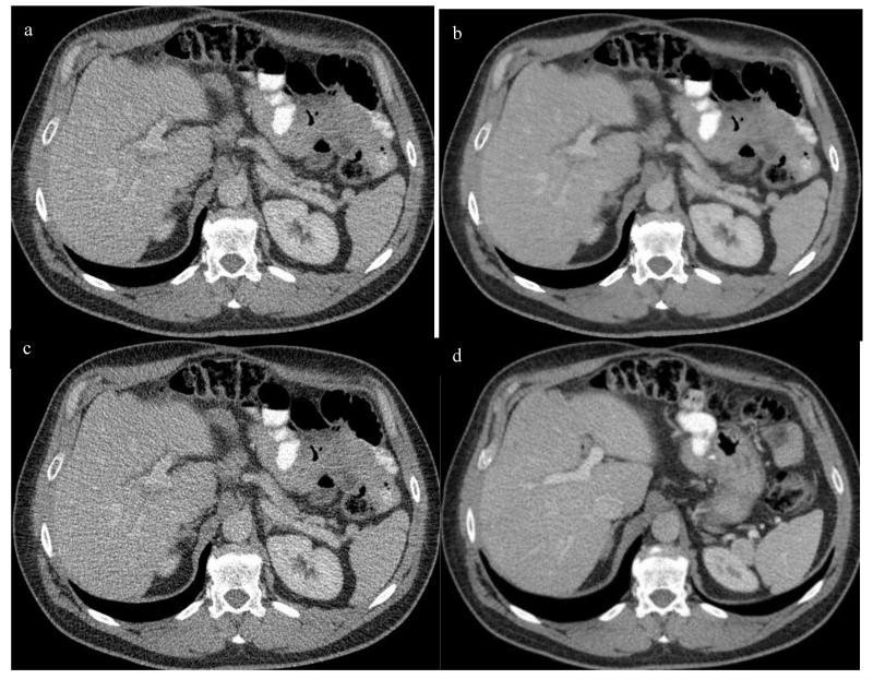
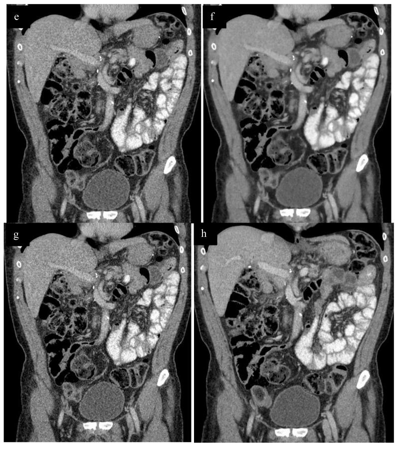
Figure 2.
Axial contrast enhanced CT in portal venous phase at the level of the main portal vein, with reduced dose series reconstructed with ASIR (a), PICCS (b), FBP (c) and standard dose FBP (d). Corresponding coronal images at the level of the portal vein are also shown (e, RD ASIR; f, RD PICCS; g, RD FBP; h, SD FBP). Images were obtained in a 54 yr old male with a BMI of 31.8 and an effective diameter of 30.4. SSDE for the reduced dose series was 5.3 mGy, compared to 13.4 mGy for the standard dose series, with an approximate dose reduction of 60% in this obese patient.


