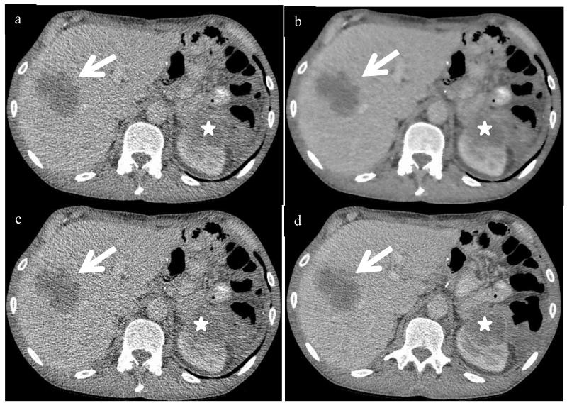
Figure 5.
Contrast enhanced axial CT images of the liver in a 52 yr old male patient s/p distal pancreatectomy and splenectomy for pancreatic adenocarcinoma with a BMI of 22 and an effective diameter of 24.9. Reduced dose images reconstructed with ASIR (a), PICCS (b), FBP (c) demonstrate an ill-defined low attenuation liver lesion which is identifiable on all series (arrows) and redemonstrated on the standard dose FBP image (d). This lesion measures roughly 50 HU compared to the background liver (95 HU) and is large enough to be well seen, but as lesions decrease in size and become closer in attenuation to background, they become more difficult to see. Note the low attenuation lesion at the upper pole of the left kidney along the post-operative bed (*) that starts to blend in to the adjacent low attenuation bowel on the reduced dose series. Both lesions were metastatic pancreas cancer. The SSDE in the reduced dose series was 2.1 mGy, compared to the standard dose series of 7.0 mGy, for a dose reduction of 70%.

