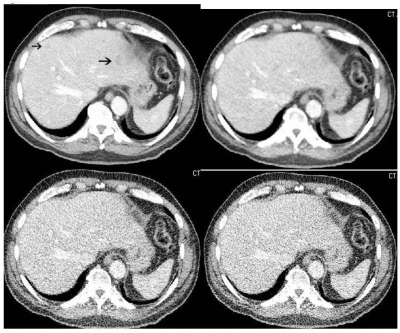
Figure 6.
Contrast enhanced axial CT images in an 82 year old male with a BMI of 28 and hepatic metastatic neuroendocrine tumor. SD-FBP image (a) demonstrates two vague low attenuation lesions (arrows) that are not well seen on the LD-PICCS (b), LD-ASIR (c) or LD-FBP (d) images. Multiple additional liver lesions (not shown here) were also not well seen on the low dose images. The SD DLP was 433, the LD DLP was 133 for a dose reduction of approximately 75%.

