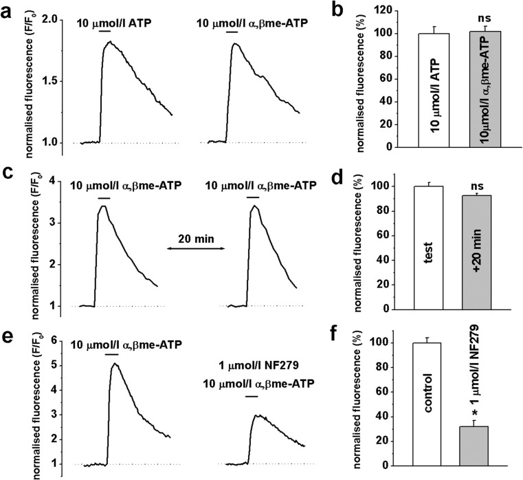
Fig. 3.
Agonist-induced changes in [Ca2+]i in HGOA VSMCs. Panel a shows representative traces of fluo-3 normalised fluorescence evoked by 3-s application of 10 μmol/l ATP and 10 μmol/l αβ-meATP. b Summarised data showed no significant difference in the effect of both agonists (n = 6, p > 0.05). c Repetitive application of 10 μmol/l αβ-meATP showed a nearly complete recovery of the agonist-induced [Ca2+]i response within 20-min interval (summary is shown in d; n = 6, p > 0.05). e Original traces of the normalised fluorescence showing that 1 μmol/l of potent P2X1 receptor blocker NF279 significantly decreased agonist-induced [Ca2+]i responses. f Summary of the mean amplitude of the normalised fluorescence in control and in the presence of the blocker (n = 6, p < 0.05, indicated by *; ns not significant

