
Figure 2. Schematic representation of GLP-1R-ir distribution in the NHP forebrain.
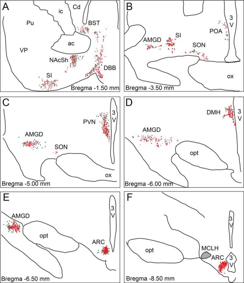
Depiction of GLP-1R-ir cell bodies (green dots) and GLP-1R-ir fibers (red lines) in forebrain sections arranged from rostral (A) to caudal (F). Gray shaded area in the magnocellular nucleus of the lateral hypothalamus (MCLH) indicates dense fiber network. 3V, third ventricle; ac, anterior commissure; BST, bed nucleus of the stria terminalis; Cd, caudate; DBB, diagonal band of Broca; DMH, dorsomedial nucleus of the hypothalamus; ic, internal capsule; NAcSh, nucleus accumbens shell; opt, optic tract; ox, optic chiasm; Pu, putamen; SI, substantia innominata; SON, supraoptic nucleus; VP, ventral pallidum.