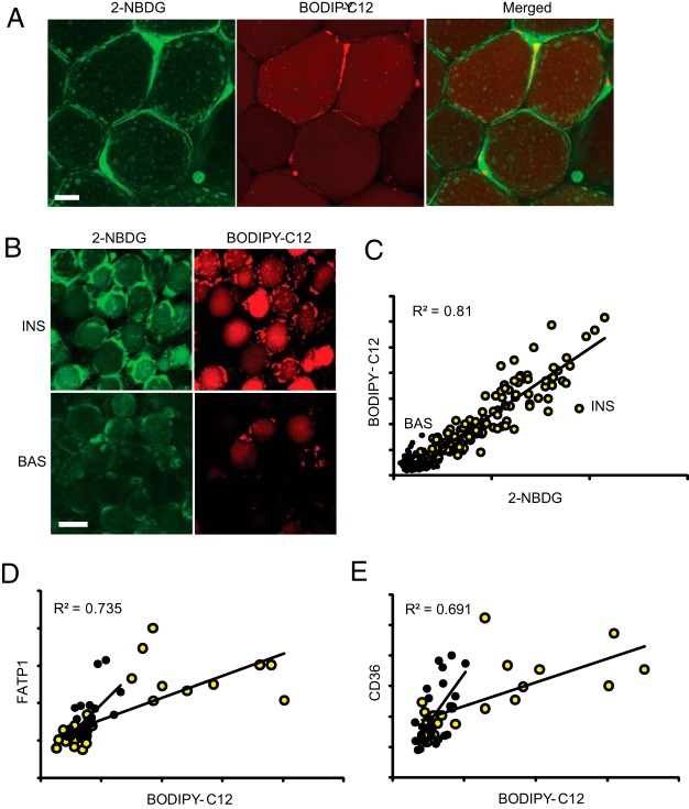
Figure 2. WAT mosaicism is related to heterogeneity in nutrient transporters.
Omental WAT explants were incubated for 2 hours in basal media (BAS) or with 10 nM insulin (INS), labeled with red BODIPY-C12 for 15 minutes, and chased in BODIPY-free media for 120 minutes in the absence or presence of insulin, followed by incubation with 2-NBDG for 1 hour, as described in Materials and Methods. A, Enlarged images show the sites of BODIPY-C12 and 2-NBDG couptake (see Results for details). B, Representative images show insulin stimulation of BODIPY-C12 and 2-NBDG uptake. Scale bar, 50 μm. C, Quantification of the total intracellular fluorescence for BODIPY-C12 and 2-NBDG. Yellow circles, insulin treated; black circles, basal. Red and green channels show significant correlation under insulin-stimulated conditions (P < .0001). D and E, Indirect immunofluorescence of BODIPY-C12-labeled WAT explants with antibodies to FATP-1 and CD36. Total BODIPY-C12, 2-NBDG, and immunofluorescent signals per cell are shown in arbitrary units.

