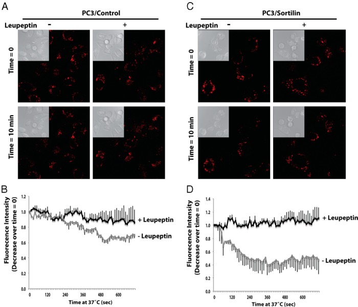
Figure 6. Progranulin levels are stabilized by lysosomal inhibition.
Serum starved PC3/Control (A and B) and PC3/Sortilin (C and D) cells were incubated with Alexa Fluor 594-labeled recombinant progranulin with or without leupeptin on ice for 1 hour. After washings, cells were shifted to 37°C and imaged at the indicated time points. These images were also acquired every 12 seconds for 12 minutes using confocal microscopy and Zeiss AIM (Supplemental Movies 1 and 2). A and C, Insets are light microscopy views of the same areas of fluorescence images. B and D, The background-corrected Alexa Fluor 594 fluorescence intensity relative to time 0 was quantified and plotted using the software Zeiss AIM 4.2 SP1. It is expressed as a function of time from experiments as in A and C, respectively. Bars are SD for n = 4.

