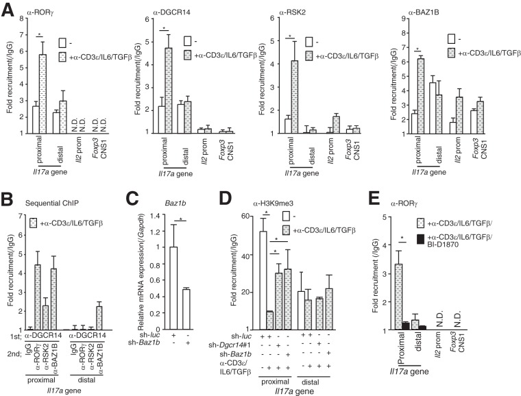
FIG 5.
ChIP analysis of the mIl17a promoter. (A) ChIP analysis of the Il17a promoter with anti-RORγ, anti-RSK2, anti-BAZ1B, and anti-DGCR14 antibodies in 68-41 cells. After cultivation of cells that were treated with or without anti-CD3ε antibody, IL-6, and TGF-β, cells were lysed and ChIP analyses were performed with the antibodies indicated. qPCR was performed with the primers indicated (see Table S3 in the supplemental material). (B) Sequential ChIP analysis of the mIl17a promoter in 68-41 cells treated with anti-CD3ε antibody, IL-6, and TGF-β. After immunoprecipitation with anti-DGCR14 antibodies, proteins were eluted with 10 mM DTT, ChIP analysis was conducted with the antibodies indicated, and qPCR was performed. The Il2 promoter and Foxp3 CNS1 were also examined, but a signal was not detected (data not shown). (C) RT-qPCR analysis of Baz1b in 68-41 cells transduced with the control (sh-luc)- or Baz1b shRNA (sh-Baz1b)-expressing retroviruses used in panel D. After transduction with each virus, GFP-positive cells were isolated with a FACS Aria (BD) and cultured. (D) ChIP analysis of the mIl17a promoter in sh-luc-, sh-Dgcr14#1-, or sh-Baz1b-expressing 68-41 cells. After cultivation with or without anti-CD3ε antibody, IL-6, and TGF-β, cells were lysed and ChIP analysis was performed with the anti-histoneH3K9Me3 antibody. A quantitative PCR assay was then performed with each primer. (E) ChIP analysis of the mIl17a promoter in 68-41 cells treated with anti-CD3ε antibody, IL-6, and TGF-β with or without BI-D1870. Cells were lysed, and ChIP analysis was performed with anti-RORγ antibody. Each experiment was performed at least three times, and results are presented as means ± SD. *, P < 0.05. N.D., not determined.

